THE OLIVE LABORATORY
  • Home
  • Science
    • Olive Lab Research
    • Pancreatic Cancer
    • Mouse Hospital
    • The KPC Model
    • Imaging
  • Lab Members
    • Alumni
  • Publications
  • More
    • A non-technical overview >
      • The Pancreas (Overview)
      • Pre-cancerous lesions (Overview)
      • Pancreatic Cancer (Overview)
      • Pancreatic Cancer Statistics
      • Symptoms and Diagnosis
      • Surgery for Pancreatic Cancer (Overview)
      • Drug Treatment (Overview)
      • Mouse Models (Overview)
      • Clinical Trials
    • Donate
    • Facilities >
      • ICRC Building
      • Shared Resources
      • Main Lab
      • Vevo 3100 Ultrasound
    • Join Us!
    • Oncology Precision Therapeutics Core >
      • MRI
      • Ivis Optical Imager
      • Vevo 3100 Ultrasound
      • Perkin-Elmer FX micro CT
    • Photos >
      • 10/30/19 Fall Fun
      • 6/29/18 Jaime's Last Day
      • 9/26/15 Mike's Wedding
      • 11/8/14 Carmine's Wedding
      • 7/7/14 Barbara's Temporary Goodbye Lunch
      • 4/26/13 Korean and Karaoke
      • 4/12/13 Purple Stride
      • 11/26/12 Pancreatic Cancer Awareness Week!!
      • 10/26/12 Pumpkin Carving
      • 9/5-7/2012 Olive and Kimmelman Lab Retreat
      • 7/13/12 Mid-summer BBQ
      • 3/29/12 Jennifer's Goodbye - BoardGame Night
      • 3/8/2012 Barbara's engagement celebration
      • 3/2/2012 Bowling
      • 12/18/2011 Holiday Party
      • 12/9/2011 Poker
      • 11/11/2011 Karaoke
      • 7/15/2011 Holiday Party
    • Support

Perkin-Elmer FX micro CT

Picture
http://www.perkinelmer.com/product/quantum-gx-microct-system-cls140083
Picture
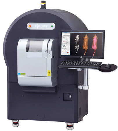
Computed Tomography (CT) is a widely used imaging modality in the clinical setting.  A CT scan makes use of computer-processed combinations of many X-ray images taken from different angles to produce cross-sectional (tomographic) images of specific areas of a scanned object, allowing the user to see inside the object without cutting. Digital geometry processing is used to generate a three-dimensional image of the inside of the object from a large series of two-dimensional radiographic images taken around a single axis of rotation.  Cross-sectional images are used for diagnostic and therapeutic purposes in various medical disciplines. Many different investigators in multiple disciplines can benefit from the use of CT images. The Columbia University Medical Center is proud to have the Perkin-Elmer Quantum FX micro CT imaging system for preclinical small animal imaging.
Powered by Create your own unique website with customizable templates.
  • Home
  • Science
    • Olive Lab Research
    • Pancreatic Cancer
    • Mouse Hospital
    • The KPC Model
    • Imaging
  • Lab Members
    • Alumni
  • Publications
  • More
    • A non-technical overview >
      • The Pancreas (Overview)
      • Pre-cancerous lesions (Overview)
      • Pancreatic Cancer (Overview)
      • Pancreatic Cancer Statistics
      • Symptoms and Diagnosis
      • Surgery for Pancreatic Cancer (Overview)
      • Drug Treatment (Overview)
      • Mouse Models (Overview)
      • Clinical Trials
    • Donate
    • Facilities >
      • ICRC Building
      • Shared Resources
      • Main Lab
      • Vevo 3100 Ultrasound
    • Join Us!
    • Oncology Precision Therapeutics Core >
      • MRI
      • Ivis Optical Imager
      • Vevo 3100 Ultrasound
      • Perkin-Elmer FX micro CT
    • Photos >
      • 10/30/19 Fall Fun
      • 6/29/18 Jaime's Last Day
      • 9/26/15 Mike's Wedding
      • 11/8/14 Carmine's Wedding
      • 7/7/14 Barbara's Temporary Goodbye Lunch
      • 4/26/13 Korean and Karaoke
      • 4/12/13 Purple Stride
      • 11/26/12 Pancreatic Cancer Awareness Week!!
      • 10/26/12 Pumpkin Carving
      • 9/5-7/2012 Olive and Kimmelman Lab Retreat
      • 7/13/12 Mid-summer BBQ
      • 3/29/12 Jennifer's Goodbye - BoardGame Night
      • 3/8/2012 Barbara's engagement celebration
      • 3/2/2012 Bowling
      • 12/18/2011 Holiday Party
      • 12/9/2011 Poker
      • 11/11/2011 Karaoke
      • 7/15/2011 Holiday Party
    • Support