THE OLIVE LABORATORY
  • Home
  • Science
    • Olive Lab Research
    • Pancreatic Cancer
    • Mouse Hospital
    • The KPC Model
    • Imaging
  • Lab Members
    • Alumni
  • Publications
  • More
    • A non-technical overview >
      • The Pancreas (Overview)
      • Pre-cancerous lesions (Overview)
      • Pancreatic Cancer (Overview)
      • Pancreatic Cancer Statistics
      • Symptoms and Diagnosis
      • Surgery for Pancreatic Cancer (Overview)
      • Drug Treatment (Overview)
      • Mouse Models (Overview)
      • Clinical Trials
    • Donate
    • Facilities >
      • ICRC Building
      • Shared Resources
      • Main Lab
      • Vevo 3100 Ultrasound
    • Join Us!
    • Oncology Precision Therapeutics Core >
      • MRI
      • Ivis Optical Imager
      • Vevo 3100 Ultrasound
      • Perkin-Elmer FX micro CT
    • Photos >
      • 10/30/19 Fall Fun
      • 6/29/18 Jaime's Last Day
      • 9/26/15 Mike's Wedding
      • 11/8/14 Carmine's Wedding
      • 7/7/14 Barbara's Temporary Goodbye Lunch
      • 4/26/13 Korean and Karaoke
      • 4/12/13 Purple Stride
      • 11/26/12 Pancreatic Cancer Awareness Week!!
      • 10/26/12 Pumpkin Carving
      • 9/5-7/2012 Olive and Kimmelman Lab Retreat
      • 7/13/12 Mid-summer BBQ
      • 3/29/12 Jennifer's Goodbye - BoardGame Night
      • 3/8/2012 Barbara's engagement celebration
      • 3/2/2012 Bowling
      • 12/18/2011 Holiday Party
      • 12/9/2011 Poker
      • 11/11/2011 Karaoke
      • 7/15/2011 Holiday Party
    • Support

Imaging

Picture
Picture

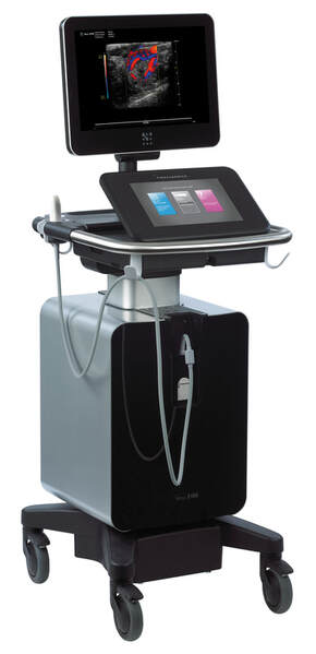
Our group utilizes high resolution ultrasound in order to detect, monitor, and quantify pancreatic tumors in mice. Using the upgraded VisualSonical Vevo 3100 high resolution ultrasound instrument, we can detect nascent tumors and premalignant lesions as small as half a millimeter in diameter.
Picture
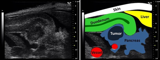
On the left is an ultrasound image of the head of the pancreas of a mouse with a 4mm diameter tumor. On the right is a color-coded map of the anatomical features in this image. Key: white= skin; green= proximal duodenum; yellow- liver; red= large blood vessels; light blue= normal pancreas; dark blue= pancreatic tumor.
Powered by Create your own unique website with customizable templates.
  • Home
  • Science
    • Olive Lab Research
    • Pancreatic Cancer
    • Mouse Hospital
    • The KPC Model
    • Imaging
  • Lab Members
    • Alumni
  • Publications
  • More
    • A non-technical overview >
      • The Pancreas (Overview)
      • Pre-cancerous lesions (Overview)
      • Pancreatic Cancer (Overview)
      • Pancreatic Cancer Statistics
      • Symptoms and Diagnosis
      • Surgery for Pancreatic Cancer (Overview)
      • Drug Treatment (Overview)
      • Mouse Models (Overview)
      • Clinical Trials
    • Donate
    • Facilities >
      • ICRC Building
      • Shared Resources
      • Main Lab
      • Vevo 3100 Ultrasound
    • Join Us!
    • Oncology Precision Therapeutics Core >
      • MRI
      • Ivis Optical Imager
      • Vevo 3100 Ultrasound
      • Perkin-Elmer FX micro CT
    • Photos >
      • 10/30/19 Fall Fun
      • 6/29/18 Jaime's Last Day
      • 9/26/15 Mike's Wedding
      • 11/8/14 Carmine's Wedding
      • 7/7/14 Barbara's Temporary Goodbye Lunch
      • 4/26/13 Korean and Karaoke
      • 4/12/13 Purple Stride
      • 11/26/12 Pancreatic Cancer Awareness Week!!
      • 10/26/12 Pumpkin Carving
      • 9/5-7/2012 Olive and Kimmelman Lab Retreat
      • 7/13/12 Mid-summer BBQ
      • 3/29/12 Jennifer's Goodbye - BoardGame Night
      • 3/8/2012 Barbara's engagement celebration
      • 3/2/2012 Bowling
      • 12/18/2011 Holiday Party
      • 12/9/2011 Poker
      • 11/11/2011 Karaoke
      • 7/15/2011 Holiday Party
    • Support